Proteins are unique molecules; they read the code of life in genes, and their own codes are also hidden in genes. Therefore, they are called the architects, pillars, and workhorses of life. A vast majority of proteins require a well-defined three-dimensional shape and flexibility for their specific and regulated function inside the cell. The hierarchy of protein structure starts with primary structure, consisting of individual amino acid residues, which then organizes itself into helices or sheets to form secondary structures. The great Prof. G. N. Ramachandran was the first to codify the geometry of the secondary structural elements in the form of backbone torsion angles. The iconic plot is called the Ramachandran Plot. Proteins further fold these secondary structural elements into tertiary and quaternary structures.
The process of formation of these structures, called Protein Folding, starts immediately at their birthplace, called Ribosomes, and most proteins emerge as perfectly folded native structures at the end of their complete synthesis. However, there are a few that require helper protein molecules called chaperones to correctly shape them. Different kinds of protein have different lifespans, and at the end of it, they are degraded to individual units with the help of another set of protein molecules, the process called Protein Unfolding. Both folding and unfolding processes are very specific, rapid, and follow a well-defined path. The Levinthal Paradox, described by Prof. Cyrus Levinthal, concluded that had it been a random search process among all possible conformations, it would take an astronomical age to fold a protein. Any aberration in folding-unfolding processes leads to dysregulation and sometimes formation of the aggregated state, resulting in disease conditions such as Alzheimer’s and Parkinson’s disease.
Prof. Christian B. Anfinsen had used ribonuclease A as a model and showed that the primary sequence of the protein under a particular set of environmental conditions (temperature, solvent concentration, and composition, etc.) at which folding occurs, dictates the formation of the native structure, which is a unique, stable, and kinetically accessible minimum of the free energy. Several studies have demonstrated that proteins unfold under different physical and chemical conditions in a test tube and refold back to their native conformation upon removal of such conditions. This helped the experimentalists to follow folding-unfolding pathways at a high spatial and time resolution and understand mechanisms. The Landscape funnel model of protein folding is currently a widely accepted model that describes a protein’s journey through the crests and troughs of internal free energy and degrees of freedom to reach the folded state from a rapidly exchanging ensemble. Proteins also take a trek through the funnel during their unfolding.
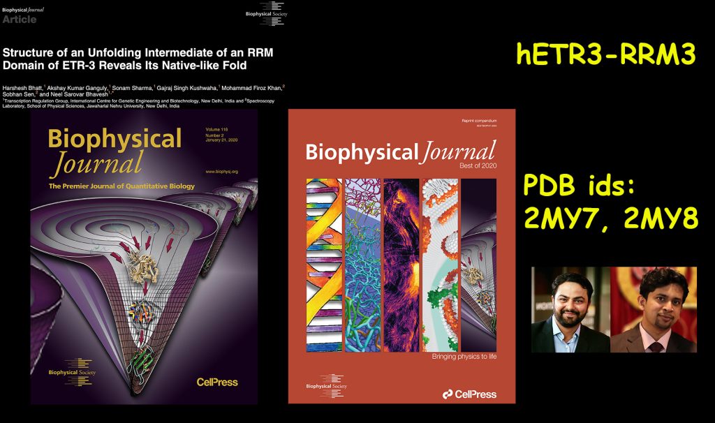
The key to understanding protein (un)folding mechanism, which is still a challenge, is high-resolution structural characterisation of all states along the funnel. In this regard, solution-state NMR spectroscopy is unparalleled in providing atomic-resolution structural and dynamics information of all the states, folded, unfolded, and intermediates, including invisibles, along the funnel. In this work, we have used a canonical RNA recognition motif (RRM) from the ETR3 protein (involved in muscular dystrophy disease) to understand the unfolding mechanism of RRM-containing protein. This is important as a similar motif in TDP-43 protein aggregates, eventually leading to neuromuscular disease conditions due to the formation of non-native structural elements. We determined the atomic-resolution structure and dynamics of folded native state (at the bottom of the funnel) and an unfolding intermediate at the middle of the funnel, and performed structural and dynamics characterisation of the unfolded ensemble rapidly dancing at the top of the funnel. Our data indicates that the intermediate state has a folded-like structure but is swollen and dynamic. It allows the solvent to access its core more easily than the folded structure. The edges of secondary structural elements are the initiation sites of unfolding. Although the unfolded state is very dynamic and lacks any structural elements, it still shows a bias for certain structural propensities, which appear to keep a memory of their folded state.
We believe that the atomic-resolution characterisation of the unfolding pathway of a canonical RNA recognition motif is likely to help understand the unfolding events in other RRMs involved in disease-causing conditions upon misfolding.
The study might also help understand those systems where protein is refolded from the purified inclusion bodies. In those cases, there could be the presence of a minor population of folded-like but not fully folded native structure.
The work was primarily done by Dr. Harshesh Bhatt and Dr. Akshay Kumar Ganguly, and the FCS work was performed in collaboration with Dr. Sobhan Sen, School of Physical Sciences, Jawaharlal Nehru University (JNU), New Delhi. The cover art was designed by Mr. Anupam Patra, a Ph.D. student.
प्रोटीन अद्वितीय अणु हैं; वे जीन में जीवन का कोड पढ़ते हैं और उनके स्वयं के कोड भी जीन में छिपे होते हैं। इसलिए, उन्हें जीवन के वास्तुकार, स्तंभ और कार्यक्षेत्र कहा जाता है। अधिकतर प्रोटीन को कोशिका के अंदर उनके विशिष्ट और विनियमित कार्य के लिए एक त्रिआयामी संरचना और लचीलेपन की आवश्यकता होती है। प्रोटीन संरचना का पदानुक्रम प्राथमिक संरचना से शुरू होता है जिसमें अमीनो एसिड के अवशेष होते हैं, जो तब द्वितीयक संरचनाओं के निर्माण के लिए हेलिकॉप्टर या शीट में व्यवस्थित होते हैं। महान वैज्ञानिक प्रो. जी. एन. रामचंद्रन ने प्रोटीन की पृष्ठ के द्वितल कोणों के माध्यम से संरचनात्मक तत्वों की ज्यामिति को संहिताबद्ध करने का कार्य किया था। इन कोणों के ऊपर उनके द्वारा बनाया गया द्वि-आयामी ज्यामिति क्षेत्र को ‘रामचंद्रन प्लॉट’ कहा जाता है। प्रोटीन इन द्वितीयक संरचनाों को तृतीयक और चतुर्धातुक संरचनाओं में बदल कर मूल संरचना का निर्माण करते हैं।
इन संरचनाओं के निर्माण की प्रक्रिया, जिसे प्रोटीन फोल्डिंग कहा जाता है, उनके जन्म स्थान पर शुरू होती है जिसे राइबोजोम कहा जाता है और अधिकांश प्रोटीन अपने पूर्ण संश्लेषण के अंत में पूरी तरह से आकृत हो मूल संरचना के रूप में उभरते हैं। हालांकि, कुछ ऐसे हैं जिन्हें जिन्हें सही ढंग से आकार पाने के लिए सहायक प्रोटीन अणुओं की आवश्यकता होती है, जिन्हे चैपरोन कहा जाता है। विभिन्न प्रकार के प्रोटीन का अलग-अलग जीवन काल होता है और इसके अंत में इन्हे एक अलग प्रकार के प्रोटीन अणुओं की आवश्यकता होती है जो उन्हें व्यक्तिगत इकाइयों अलग-अलग कर देता है, इस प्रक्रिया को प्रोटीन अनफोल्डिंग कहते है। फोल्डिंग और अनफोल्डिंग दोनों प्रक्रियाएं बहुत विशिष्ट, तीव्र और एक परिभाषित पथ का अनुसरण करती हैं। प्रो. साइरस लेविंथल द्वारा वर्णित लेविथल पैराडॉक्स के अनुसार अगर प्रोटीन अपने हर एक संभावित आकृति को समन्वेषित कर अपनी सही आकृति को प्राप्त करने की चेष्टा करे तो इस प्रक्रिया को खगोलीय उम्र लग जाएगी। फोल्डिंग-अनफोल्डिंग प्रक्रियाओं में किसी भी तरह की गड़बड़ी से विकृति उत्पन्न हो जाती है और कभी-कभी इस स्थिति में प्रोटीन का ढेर बनकर द्रव से आ जाते हैं और कोशिकाओं में जम जाते हैं, जिसके परिणामस्वरूप अल्जाइमर और पार्किंसंस रोग जैसे रोग होते हैं।
प्रो. क्रिश्चियन बी. अनफिंसन ने राइबोन्यूक्लिज़ ए प्रोटीन पर प्रयोग करके ये दिखाया था कि एक विशेष पर्यावरणीय परिस्थितियों (तापमान, विलायक एकाग्रता और संरचना, आदि) के तहत प्रोटीन का प्राथमिक अनुक्रम जिस पर तह होती है, मूल संरचना के गठन को निर्धारित करता है, जो एक अद्वितीय, स्थिर और बलगति रूप से सुलभ न्यूनतम ऊर्जा वाली स्थिति है। कई अध्ययनों से पता चला है कि एक टेस्ट ट्यूब में विभिन्न भौतिक और रासायनिक स्थितियों में प्रोटीन की अपनी संरचना खुल जाती है और ऐसी स्थितियों को हटाने पर वह अपनी मूल पुष्टि को वापस प्राप्त कर लेता है। इससे प्रायोगिकों को एक उच्च स्थानिक और समय प्रमाणिकता पर फोल्ड-अनफोल्डिंग मार्ग और उसके तंत्र को समझने में मदद मिलती है। प्रोटीन फोल्डिंग का परिदृश्य कीप (लैंडस्केप फ़नल) तंत्र वर्तमान में व्यापक रूप से स्वीकृत तंत्र है, जो तीव्र मुक्त ऊर्जा से मुग्ध अवस्था में पहुँचने के लिए आंतरिक मुक्त ऊर्जा और स्वतंत्रता-श्रेणी के शिखाओं और गर्तों के माध्यम से प्रोटीन की यात्रा का वर्णन करता है। प्रोटीन अपने संरचना खुलने के दौरान कीप के माध्यम से ही यात्रा करते हैं।
प्रोटीन की संरचना के बनने और खुलने के तंत्र को समझने की कुंजी, जो अभी भी एक चुनौती है, कीप के भीतर के सभी संरचनाओं के उच्च-स्तर पे संरचनात्मक वर्णन अत्यावश्यक है। इस संबंध में एन.एम.आर. स्पेक्ट्रोस्कोपी सभी आकृतियों के परमाणु-स्तर की संरचनात्मक और गतिशीलता की जानकारी प्रदान करने में अद्वितीय है, और साथ ही अदृश्य, अनफोल्डेड और मध्यवर्ती सहित सभी आकृतियों की जानकारी प्रदान करता है। इस काम में हमने आर. आर. एम. युक्त प्रोटीन के अनफोलोइंग तंत्र को समझने के लिए ई. टी. आर.-३ (ETR-3) प्रोटीन (मस्कुलर डिस्ट्रॉफी रोग से जुड़ा) से एक विहित आर.एन.ए. रिकग्निशन मोटिफ (RRM) का उपयोग किया है। यह टी.डी.पी.-४३ प्रोटीन में पाए जाने वाले एक समान रूपांकनों के रूप के कारण महत्वपूर्ण है और जिसमे गैर-मूल संरचनात्मक तत्वों के गठन के कारण स्नायु-पेशी (न्यूरोमस्कुलर) रोग उत्पन्न होता है। हमने ई. टी. आर.-३ प्रोटीन के आर. आर. एम.-३ की मूल स्थिति (कीप के नीचे) और कीप के मध्य में की एक थोड़ी खुली हुई स्थिति की परमाणु-स्तर पर संरचना और गतिशीलता निर्धारित की और कीप के शीर्ष पर उन्मुक्त नृत्य करते हुए पूर्णतः खुले हुए आकृति समूह की संरचनात्मक और गतिकी की जानकारी प्राप्त की। हमारे द्वारा प्राप्त तथ्य ये बताता है कि मध्यवर्ती आकृति अपने मूल संरचना की तरह है लेकिन उसमे सूजन और अधिक गतिशीलता है। इस संरचना के अन्दर द्रव (पानी) को मूल संरचना की तुलना में अधिक आसानी से जा पाते हैं। माध्यमिक आकृति के संरचनात्मक तत्वों के किनारे प्रोटीन के खुलने के आरम्भिक स्थल हैं। हालांकि पूर्णतः खुले हुआ आकृति समूह बहुत ही गतिशील है और उसमे किसी भी संरचनात्मक तत्वों की कमी है, फिर भी यह कुछ विशेष संरचनात्मक स्थितिओं के लिए पूर्वाग्रह दिखाती है, जो उनके मूल संरचना की स्मृति रखने जैसा प्रतीत होता है।
हम मानते हैं कि हमारे द्वारा वर्णित परमाणु-स्तर की जानकारी से विभिन्न रोगों में शामिल अन्य आर. आर. एम. के संरचना खुलने की घटनाओं को समझने में मदद मिलेगी |
यह अध्ययन उन प्रणालियों को समझने में भी मदद कर सकता है जहां प्रोटीन थक्केदार अवस्था से वापस मूल संरचना में लाया जाता है। इन मामलों में मूल संरचना जैसी मामूली आबादी की भी मौजूदगी हो सकती है, जो पूरी तरह से मूल संरचना में ना हों।
यह शोध मुख्यतः डा. हर्षेश भट्ट और डा. अक्षय कुमार गांगुली द्वारा किया गया और FCS कार्य जवाहरलाल नेहरू विश्वविद्यालय (जे. एन. यू.) के भौतिक विज्ञान के डा. सोभन सेन के सहभागिता से की गई | मुख्य पृष्ठ आवरण को श्री अनुपम पात्रा, वर्तमान पी.एच.डी. छात्र, ने बनाया है।
References
- Dill K.A. and Chan H.S. (1997) From Levinthal to pathways to funnels. Nat. Struct. Biol. 4: 10-19
- Bhatt H, Ganguly AK, Sharma S, Kushwaha GS, Khan MF, Sen S and Bhavesh NS (2020) Structure of an unfolding intermediate of an RRM domain of ETR-3 reveals its native-like fold. Biophys. J. 118, 352-365.
- Structure co-ordinates: Protein Data Bank (PDB) accession numbers 2MY7 and 2MY8. Sequence-specific resonance assignments: BioMagResBank (BMRB) IDs: 19316 and 19685.